This course is aimed at nurses working in general practice / referral practice
Learning objectives
After completion of this week, participants should be able to:
Learning materials this week:
(released on Monday morning for on demand learning until the course ends – approximate timings)
Learning objectives
After completion of this week, participants should be able to:
Learning materials this week:
(released on Monday morning for on demand learning until the course ends – approximate timings)
Learning objectives
After completion of this week, participants should be able to:
Learning materials this week:
(released on Monday morning for on demand learning until the course ends – approximate timings)
Learning objectives
After completion of this week, participants should be able to:
Learning materials this week:
(released on Monday morning for on demand learning until the course ends – approximate timings)
Learning objectives
After completion of this week, participants should be able to:
Learning materials this week:
(released on Monday morning for on demand learning until the course ends – approximate timings)
Learning objectives
After completion of this week, participants should be able to:
Learning materials this week:
(released on Monday morning for on demand learning until the course ends – approximate timings)
This course will be fully tutored by Ash Moors and will consist of 15 hours of CPD given in various formats, including tutorials, tasks, case studies, forum discussions and quizzes. This course is tutored for 6 weeks, followed by a two week extension of untutored ‘catch up’ time, before the course officially ends.
All delegates will then have unlimited lifetime access to the learning material for future reference

Ash Moors FdSc, GradDipVN, PGCertVedEd, FHEA, RVN
Radiographer, Royal Veterinary College
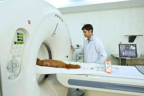
Ash graduated from the Royal Veterinary College with an FdSc in Veterinary Nursing in 2010, and worked in first opinion Practice in Dorset with access to low field MRI and digital radiography systems. He returned to the RVC in 2012, undertaking the Graduate Diploma in Clinical and Professional Veterinary Nursing whilst continuing to work in first opinion practice, choosing to undertake the surgical nursing and diagnostic imaging elective modules, which further developed his interest in radiography. During this period he built and coded a custom image storage system for the practice he worked at, allowing query and retrieval of digital imaging studies.
This online course is worth 15 hours of CPD.
A certificate will be available from the ‘My Courses’ section, for you to download and print, once you have completed the course. A permanent record of your total CPD hours will also be recorded in your account section.
The Australian Veterinary Nurse and Technician (AVNAT) Regulatory Council has allocated 15 AVNAT CPD points to this continuing education activity.
This course is also recognised by the New Zealand Veterinary Nursing Association (NZVNA) as providing 15 CPD points.
Upon purchase you will be registered to attend the course, for 6 weeks from the start date. The course will consist of various interactive tasks and lessons, including quizzes, case studies, forum discussions and further reading material.
The course is fully tutored, with new material will be provided each Monday morning, but the onus will be on the individual delegate to ensure that all tasks are fully complete. The certificate will only be issued at the end of the course when all tasks have been accomplished. Fewer CPD hours will be awarded at the the end of the course if there are unfinished tasks or there has been no contribution to the discussion forum, for example
The course is fully flexible, and there are no weekly ‘deadlines’ – the lessons and tasks may be completed whenever is convenient for each delegate, and any live lessons with be recorded and made available later that same day. Furthermore, all the course material will be available for a further 2 weeks, to allow delegates the opportunity to catch up on missed lessons and tasks, or to take the opportunity to delve further into the suggested reading texts. Please note however, that the course will not be tutored by the speaker during these final two weeks, but the time spent will count towards your CPD hours
After 8 weeks, the course will be complete and there will be no further opportunity to gain the certificate or CPD hours, however, you will have unlimited lifetime access to the tutorials, further reading and quizzes for future reference. If you make any personal notes during the course using the ‘take notes’ app, these will be saved, along with your certificate and CPD record for permanent access in ‘My CPD’
This course has been listed as ‘Intermediate / Advanced’ level
All of our courses are aimed at veterinary nurses in general practice, but everyone who works in the veterinary profession is very welcome to attend, whether you are a clinical receptionist, veterinary surgeon, student nurse or have been a qualified nurse for over 20 years!
The courses are not formally assessed for skill level, so the following CPD levels are just a rough guide to help you decide if a course may be more or less suitable:
Introduction
– maybe most suitable for qualified nurses in general practice approaching a new topic or looking for a refresher course
Intermediate
– maybe most suitable for qualified nurses in general practice along with referral / specialist nurses looking for a refresher course
Advanced
– maybe most suitable for referral / specialist nurses and highly experienced qualified nurses in general practice
You will earn 255 Pawprint Points (£25.50 website credit) when you purchase this course
Our loyalty scheme rewards you with 10% in website credit to spend on future courses. Choose pay with Pawprint Points during checkout.

Iga Sieraj –
Amazing course with great resources. Very informative webinars, learned a lot!
Rated: Advanced level
(Referral or specialist RVN – UK)
Marie Gannon –
Excellent course. Intense and highly informative. By far the best I have done.
Rated: Advanced level
(Referral or specialist RVN – UK)
Rachel Tretton –
Really enjoyed the past 6 weeks. Thank you Ash!
Rated: Intermediate / Advanced Level
(Referral or specialist RVN – UK)